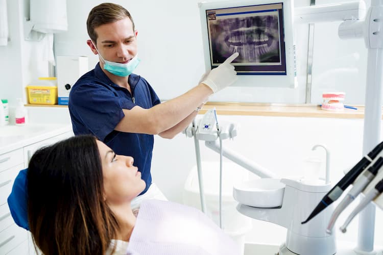
Also known as dental radiographs, dental X-rays use controlled pulses of radiation to create images of the internal structures of the jaw and mouth. Dental X-rays are useful for viewing jawbones and various tooth structures. They can find and image cavities, bone or gum loss, periodontal disease, benign or malignant tumours, and other normal or abnormal structures within the lower portion of the head. In children and adolescents, they are also useful for finding un-erupted permanent teeth and imaging root structures in preparation for orthodontic work.
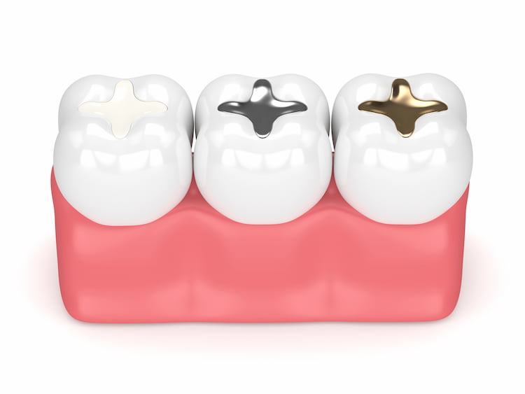
Like other types of X-rays, dental X-rays take advantage of the natural density contrasts within the mouth and jaw. For instance, denser jawbones, teeth, crowns and fillings show up as light areas within the darker, less-dense soft tissues that surround them. Cavities easily appear on X-rays because they are less dense than the teeth that they affect. Dentists, doctors and other medical professionals have utilized the relatively simple concept of X-ray imaging since the late 1800s.
Although dental X-rays use radiation to achieve light-dark contrasts, they are not dangerous when used occasionally. During a typical X-ray session, a patient receives about as much radiation exposure as he or she would on a five-hour airplane flight. Lead shields and collars further reduce these exposure levels.
